CBCT Imaging
CBCT imaging is a powerful diagnostic tool that allows for more precise, safer, and more predictable dental care by giving your provider a complete picture of your oral health.
Why Is It Needed?
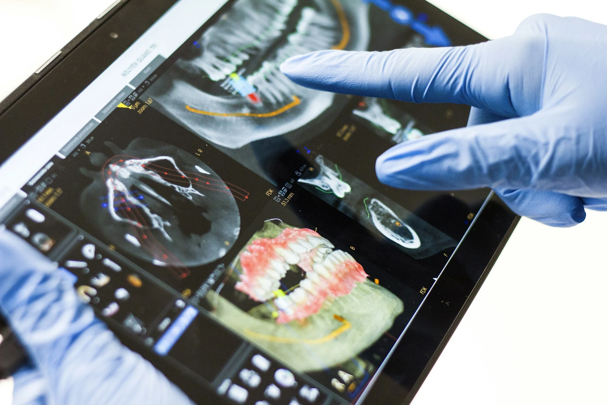
CBCT (Cone Beam Computed Tomography) imaging provides highly detailed 3D views of your teeth, jaw, nerves, and surrounding structures. Unlike traditional dental X-rays, which produce flat images, CBCT scans allow your dentist to see precise anatomical details from multiple angles.
This technology is often recommended when more advanced imaging is required, such as:
Planning dental implants
Evaluating bone structure and density
Diagnosing impacted teeth or abnormalities
Assessing jaw joint issues (TMJ)
Detecting infections, cysts, or tumors
Preparing for complex extractions or surgeries
CBCT helps ensure accurate diagnosis and treatment planning, reducing the risk of complications.
What Happens During the Procedure?
CBCT imaging is quick, non-invasive, and painless.
During the scan:
You will be seated or standing comfortably
A specialized scanner rotates around your head
The machine captures multiple images in seconds
These images are combined to create a detailed 3D model
The process typically takes less than a minute, and no special preparation is needed.
Aftercare
What to expect:
You can resume normal activities immediately
No discomfort or side effects
Your dentist will review the images and discuss findings with you
Because CBCT uses a low dose of radiation, it is considered safe, though it is only used when clinically necessary.
Benefits of CBCT Imaging
CBCT technology offers significant advantages in modern dental care:
Detailed 3D visualization of oral structures
More accurate diagnosis compared to traditional X-rays
Improved treatment planning and outcomes
Faster and more efficient imaging process
Non-invasive and comfortable for patients
Helps reduce surprises during procedures